A RARE BUT IMPORTANT CAUSE of
SYNCOPE
from Southern Medical Journal
Kirk M. Chan-Tack, MD, Department of Medicine, Hospital of the
University of Pennsylvania, Philadelphia
Abstract
An elderly woman came to
our emergency room for evaluation of a syncopal episode. While
climbing a flight of stairs, she had turned her head to the
left and abruptly passed out. Positive physical findings included
blood pressure of 141/65 mm Hg (right arm) and 80/43 mm Hg (left
arm), as well as nonpalpable left radial and brachial pulses
that were detectable only by Doppler ultrasonography. Carotid
duplex ultrasonography showed reverse flow in the left vertebral
artery and an abnormal, stenotic distal left subclavian artery.
Magnetic resonance angiography confirmed complete occlusion
of the left subclavian artery with classic subclavian steal.
The patient had percutaneous transluminal angioplasty with stenting
of the left subclavian artery and has remained asymptomatic
through 2 years of follow-up with aggressive risk-factor modification.
Introduction
Subclavian steal syndrome
is caused by occlusion of the proximal subclavian artery with
subsequent retrograde filling of the subclavian artery via the
vertebral artery. The decreased blood flow to the brain and
upper extremity on the affected side can result in an array
of symptoms, classified broadly as due to (1) vertebrobasilar
insufficiency or (2) ischemia of the affected extremity. Vertebrobasilar
insufficiency may produce light-headedness, dizziness, vertigo,
ataxia, visual disturbances, motor deficits, focal seizures,
confusion, aphasia, headache, presyncope, or syncope. Symptoms
due to ischemia of the affected extremity are less frequent
and include weakness, arm claudication, paresthesias, or coldness
on the affected side. Risk factors for this syndrome include
hypertension, diabetes, hypercholesterolemia, tobacco use, and
vigorous exercise of the affected extremity. This report describes
a patient with syncope as the presenting symptom of subclavian
steal syndrome.
Case Report
A 79-year-old woman was
admitted for evaluation of a syncopal episode. The patient was
in her usual state of health until the morning of admission
when, while climbing a flight of stairs, she turned her head
to the left and abruptly passed out. She fell down 12 stairs
and sustained a left occipital laceration. The patient denied
chest pain, palpitations, prodrome, visual changes or aura,
bowel or bladder incontinence, tongue biting, or postictal state.
She had had no previous episodes of presyncope or syncope.
Her medical history included longstanding hypertension, type
2 diabetes, hypercholesterolemia, and tobacco use. Medications
included fosinopril, repaglinide, and atorvastatin. Family history
was notable only for diabetes. She had a 40 pack-year history
of tobacco use but never used alcohol or illicit drugs. Review
of systems was otherwise negative.
On physical examination, temperature
was 37.2°C (98.9°F), blood pressure 141/65 mm Hg (right
arm), heart rate 76/min and regular, and respiratory rate 16/min.
A 6 cm laceration was present on the left occiput. No carotid
bruits were heard. The lungs were clear bilaterally. Cardiac
examination showed a regular rate and rhythm, normal S1 and
S2, and no murmurs, rubs, or gallops. Findings on abdominal
and neurologic examinations were normal. Peripheral pulses were
2+ throughout, with the notable exception of the left radial
and brachial pulses, which were detectable only by Doppler ultrasonography.
After discovery of the nonpalpable left radial and brachial
pulses, the blood pressure was measured in the left arm and
found to be markedly low at 80/43 mm Hg.
Admission complete blood count,
chemistry panel (including cardiac enzymes and troponin), electrocardiogram,
and chest radiograph were all unremarkable. Noncontrast computed
tomography of the head was negative for hemorrhage, infarct,
or mass effect. Clinical findings were highly suggestive of
subclavian steal. Carotid duplex ultrasonography showed reverse
flow in the left vertebral artery and abnormal, stenotic distal
left subclavian artery. Magnetic resonance angiography confirmed
complete occlusion of the left subclavian artery with classic
subclavian steal ( scas-Figs 1.jpg, -2.jpg, and -3.jpg).
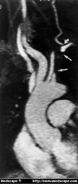
Scas-Figure 1. Magnetic
resonance angiogram of proximal great vessels shows occlusion
of left subclavian artery (arrows) with reconstitution proximal
to origin of left vertebral artery, as well as stenosis of left
vertebral artery origin (curved arrow).
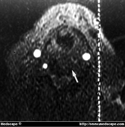
Scas-Figure 2. Magnetic
resonance time-of-flight angiogram of mid-neck (depicting superiorly
flowing blood) shows absence of superior flow in left vertebral
artery (arrow) consistent with occlusion or reversal of flow.
(Vertical bright line was artifactual.)

Scas-Figure 3. Magnetic
resonance time-of-flight angiogram of mid-neck (displaying inferiorly
flowing blood) shows reversal of flow in left vertebral artery
(arrow). (Vertical bright line was artifactual.)
Vascular surgery consultation
was obtained, and the patient had percutaneous transluminal
angioplasty (PTA) with stent placement in the left subclavian
artery. She tolerated the procedure well and was discharged
on the following day. With close monitoring and support from
her primary care physician, the patient has stopped smoking
and has also achieved excellent control of her hypertension,
diabetes, and hypercholesterolemia. She maintains a moderate
level of exercise and activity (including use of her left arm)
and has remained asymptomatic through 2 years of follow-up.
Discussion
Subclavian steal syndrome
is rare; its incidence and prevalence are unknown. It has a
2:1 male-female ratio, and patients are generally aged 55 years
or older.
Subclavian steal syndrome is caused by occlusion of the proximal
subclavian artery. Arteriosclerosis is the cause in 95% of cases.
Less common causes of occlusion include dissecting aortic arch
aneurysm, embolus, and Takayasu's arteritis. The underlying
pathophysiology of subclavian steal syndrome is the development
of a negative pressure gradient between the vertebral-basilar
and vertebral-subclavian artery junctions. Subsequent retrograde
filling of the subclavian artery via the vertebral artery causes
the subclavian artery to "steal" blood from the vertebrobasilar
system. Decreased blood flow to the brain and upper extremity
on the affected side results in a variety of symptoms, classified
broadly as due to (1) vertebrobasilar insufficiency or (2) ischemia
of the affected extremity.Vertebrobasilar insufficiency may
produce light-headedness, dizziness, vertigo, ataxia, visual
disturbances, motor deficits, focal seizures, confusion, aphasia,
headache, presyncope, or syncope. Although rare, strokes and
resulting deaths have occurred due to the subclavian steal syndrome.
Symptoms due to ischemia of the affected extremity are less
frequent and include weakness or arm claudication after exercise,
paresthesias, or coldness on the affected side. Both symptom
subtypes are classically reproducible by vigorous exercise of
the affected arm, often in conjunction with sudden or sharp
turning of the head to the affected side.
Risk factors for this syndrome
include hypertension, diabetes, hypercholesterolemia, tobacco
use, and vigorous exercise of the affected extremity, all of
which were present in this patient. Hypertension increases the
pressure in the basilar arterial system. Diabetes, hypercholesterolemia,
and smoking all worsen endothelial and vascular integrity. Vigorous
exercise of the affected extremity also decreases pressure in
the subclavian arterial system. This further increases the negative
pressure gradient between the vertebral-basilar and vertebral-subclavian
artery junctions, causing symptoms as described. If vigorous
exercise of the affected arm is combined with sudden or sharp
turning of the head to the affected side, as occurred in this
patient, the negative pressure gradient is even steeper. Physical
findings of subclavian steal syndrome include unilaterally decreased
pulses, >20 mm Hg difference in blood pressure between the
upper extremities, supraclavicular bruits, and disappearance
of the radial pulse with exercise of the affected extremity.
Although subclavian steal is
rare, a high index of suspicion is warranted in the presence
of a suggestive history, risk factors, and physical findings.
Differential diagnosis includes intracranial vascular disease,
carotid artery disease, vertebral artery disease, brain tumor,
and subdural hematoma. Diagnosis of subclavian steal syndrome
confirmed by (1) carotid duplex ultrasonography (which shows
reversal of vertebral artery flow and a stenotic distal subclavian
artery) and (2) magnetic resonance angiography or arch aortography
(which shows subclavian artery occlusion and absence of vertebral
artery flow). Risk factor modification (smoking cessation and
control of hypertension, diabetes, and hypercholesterolemia)
is essential. Patients are also educated about preventing injury
to and reducing exercise of the affected arm. Invasive treatment
of subclavian steal syndrome is necessary for symptomatic patients.
Options include axillo-axillary bypass, carotid-subclavian bypass,
and PTA of the stenotic proximal subclavian artery with stent
placement. The prognosis is excellent with all three modalities
(95% of patients remain asymptomatic after the procedure), but
PTA is preferred because of its lower morbidity, shorter hospitalization,
and faster recovery. Follow-up includes assessment for recurrence
of symptoms, blood pressure measurements in both arms, and risk
factor modification.
Conclusion
This case underscores the
importance of subclavian steal syndrome, its wide range of clinical
manifestations (including syncope), and its morbidity and potential
for mortality if undiagnosed or misdiagnosed, Recognition of
this syndrome is crucial, since patients can be successfully
treated with PTA and stent placement or other bypass graft operations.
Aggressive risk factor modification (smoking cessation and control
of hypertension, diabetes, and hypercholesterolemia) is also
essential in the treatment of subclavian steal syndrome
South Med J 94(4):445-447, 2001. ©
2001 Southern Medical Association
ILLUSTRATIONS of SUBCLAVIAN
ARTERY STEAL
from http://www.vesalius.com/graphics/archive/archtn.asp?VID=514&nrVID=22
 angoccsca-fig1.jpg
Angiogram of occluded subclavian
artery
 angoccsca-fig2.jpg
Retrograde filling of left subclavian
artery

Subclavian steal blood flow
.


