In
patients vulnerable to sustained ventricular tachycardia(VT)
may be found low amplitude, high frequency waveforms in the
terminal QRS complex of the electrocardiogram called ventricular
late potentials, which are continuous with the QRS complex and
persist for tens of milliseconds into the ST segment.
| Pathophysiological
Basis Of Late Potentials |
They
appear to arise form small areas of structurally abnormal myocardium
(heart muscle) in which ventricular activation is delayed and
asynchronous. When surviving heart fibers are separated by connective
tissue, heterogenous activation patterns may occur. The result
is a low- amplitude, fragmented local electrogram. This activity
can be recorded in most patients with remote myocardial infarction
(heart attack), but is detected at fewer recording sites and
is of shorter duration in infarction patients without clinical
VT.
Late potentials imply that the substrate for reentry is present,
and then be precipitated by such triggers as premature ventricular
beats, myocardial ischemia (lack of oxygen), electrolyte imbalance
(like low potassium), or autonomic nervous system instability.
Healthy volunteers rarely have abnormal SAECGs. Patients with
sustained, inducible monomorphic VT after myocardial infarction
have abnormal SAECGs in 79 to 92% of cases. Late potentials
occur more frequently and are of greater duration in patients
with monomorphiic sustained VT than in patients with ventricular
fibrillation, a rhythm less associated with conduction delay.
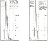
Using
a Butterworth filter multiple samples of a repeating wave form
are averaged to exclude randomly occurring noise, having excluded
ectopic ventricular beats and excessively noisy beats. After
signal averaging, the ECG is high-pass filtered to reduce the
low-frequency signals contained in the QRS complex and ST segment.This
is necessary because the repolarization phases of the action
potential produces slowly changing and lower frequency signals
that interfer with the measurement of microvolt signals corresponding
to the delayed depolarization of small areas of myocardium (figure
143).
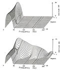
Frequency
domain analysis using a Fourier transform is another means to
extract diagnostic high frequency content from the SAECG (figure144).
The analysis of multiple segments may allow better separation
between noise and late potential.
| VENTRICULAR
LATE POTENTIALS |
Common
criteria defining of a late potential include the following:
1) the filtered QRS complex is longer than 114ms,
2) the terminal filtered QRS complex remains below 40 microvolt
for more than 38ms, and
3) there is less than 20microvolt of signal in the last 40ms
of the filtered QRS complex.
Late
potentials are not present universally in patients with recurrent
VT. In some instances the fragmented activity may be too brief
or the late potential may be masked by bundle branch block.
The signal amplitude may be too low to be differentiated from
noise. Delayed fragmented electrical activity is probably related
to reentry ventricular arrhythmias. Other potential electrophysiologic
mechanisms of VT, such as increased automaticity or triggere
activity, may not be associated with late potentials. Patients
with VT in the absence of structural heart disease rarely have
late potentials.
Prognostic
Value of late potentials after acute myocardial infarction.
Several
studies in post myocardial infarction patients have shown an
increased likelihood of spontaneous VT or sudden cardiac death
in patients who have an abnormal SAECG. Abnormal SAECGs are
found in 26 to 40% of postmyocardial infarction patients when
the recording is made prior to hospital discharge.
Fourteen to 29% of patients recovering from myocardial infarction
with abnormal SAECGs will experience sustained VT during the
first year, compared to 0.8 to 4.5% of those with a normal recording.
A majority of patients who have an abnormal SAECG do not develop
a serious arrhythmia.
The negative SAECG coupled with normal left ventricular function
suggests less need for concern about arrhythmias and less need
for ambulatory monitoring and drug therapy of of ventricular
ectopy. Patients with abnormal SAECGs are more prone to inducible,
sustained monomorphic VT, ventricular fibrillation, or sudden
death.
Reference:Walter,P.,MD,Hurst's
The Heart,8th Edition,Technique of Signal- Averaged Electrocardiography,pp.893-896.