


Magnetic Resonance Imaging
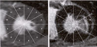
Magnetic resonance imaging (MRI) is a technique for obtaining high resolution images of various organs within the human body by mapping the distribution of hydrogen nuclei.
The patient is placed in a strong external magnetic field, causing the atomic nuclei of the part of the body being studied to line up and create a magnetic field. These nuclei are exposed to a radiofrequency (RF) electromagnetic wave, and absorb energy from it. This absorption of energy is called resonance.
When the RF wave stops, the atoms relax back to equilibrium and release the absorbed energy to the environment as RF wave emissions, which can be detected with sensors. To create a two dimensional picture, two magnetic field gradients are applied to the organ to be studied, one perpendicular to the other. The RF is applied after each magnetic field. A computer detects the RF emission and reconstructs it into a picture (figures 23b, 122, 123, 124, 125 ).
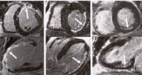
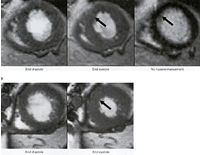
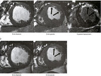
Recent studies have shown that MRI after the intravenous administration
of a gadolinium-based contrast agent such as gadopentetate dimeglumine
can distinguish between reversible and irreversible myocardial
ischemic injury, regardless of the extent of the resultant wall
motion abnormality or the age of the infarct or heart attack
(see illustrations).
These results can be used to predict whether regions of abnormal
ventricular contraction due to the infarct will improve after
revascularization (bypass surgery or angioplasty ) in patients
with coronary artery disease.
It has been shown that areas of the myocardium which are hyperenchanced
are nonviable and do not recover to contract normally after
revascularization (figures 124, 125 ). While areas which do
not hyperenhance from the gadolinium injection are viable and
do recover and contraction improves after revascularization.
The advantage over other methods like echocardiography and positron emission tomography is that the hyperenhanced MRI (due to high spatial resolution) shows the transmural extent of viable myocardium with the nonviable myocardium being highlighted and has greater accuracy in muscle segments with the most severe dysfunction (while nuclear scintigraphy and echocardiography have reduced predictive accuracy if more severe dysfunction of the myocardial contractionis present).