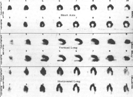
Figure 109h
Tomographic slices two pixels in thickness form a thallium stress and 4-h redistribution study in standard display. There is an inferoposterior defect on the initial tomographic slices that shows almost complete redistribution at 4 h. This scan was read as showing ischemia in the distribution of the RCA.
Johnson, L.L., MD, Pohost, G.M., MD, Nuclear Cardiology, Hursts The Heart, 8th edition, p 2281-2295.