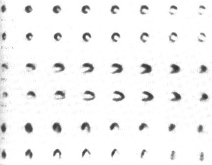
Figure 109i
Tomographic slices one pixel thick from a thallium stress and 4-h redistribution study. The vertical long axis slices are displayed from lateral wall to septum and the horizontal long axis slices are displayed from superior to inferior walls. There is an inferiolateral defect on the initial tomographic slices that shows minimal redistribution at 4 h and no further change following thallium reinjection (not shown). This scan was read as showing infarction in the distribution of the LCF artery.
Johnson, L.L., MD, Pohost, G.M., MD, Nuclear Cardiology, Hursts The Heart, 8th edition, p 2281-2295.