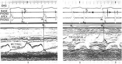
Figure 198a
The apex phonocardiography is recorded with the mitral valve echocardiogram (left panel) and tricuspid valve echocardiogram (right panel) in a normal subject. The mitral (M1) and tricuspid T1 components of the first heart sound are coincident with the closure points C. of the mitral and tricuspid valves respectivel. A small low frequency vibration (m) is seen prior M1, and a few low frequency vibrations follow T1 in early systole.