Figure 199g
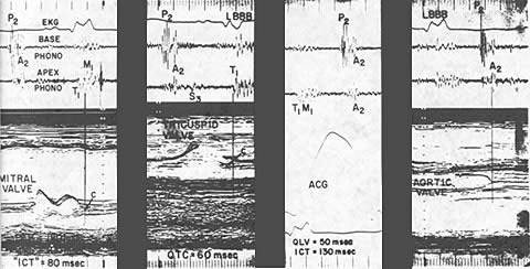
Base and apex phonocardiograms are recorded simultaneously with the mitral and tricuspid valve echocardiogram, the apex cardiogram (ACG), and the aortic echocardiogram in a patient with LBBB and significant left ventricular dysfunction. A markedly delayed M1 is coincident with complete coaptation of the anterior and injury and posterior leaflets of the mitral valve. The sequence of S1 is reversed. The mitral valve echocardiogram shows significant atriogenic preclosure of the anterior and posterior leaflets and is followed by a slow ventricular closure of the valve, resulting in a soft the M1. in the next panel, T1 is clearly shown to be coincident with final closure point of the tricuspid valve and precedes M1 .Reversed splitting of S2 is present and is confirmed by the simultaneous recording of the aortic valve echocardiogram in the far right panel. In this patient with LBBB, the electromechanical interval ( Q-left ventricular) was within normal limits, and the reversed splitting of S1 andS2 to was due to severe left ventricular dysfunction with mark prolongation of isovolumic contraction time.