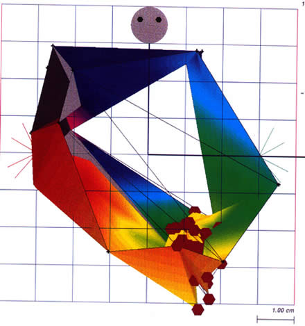
Figure 206-3
(Plate 75) Anterior-posterior view of the right atrium during typical, inferior vena cava (IVC)tricuspid valve annulus isthmus-dependent atrial flutter using the Biosense CARlO system. The red shows the earliest activation with respect to the timing reference (typically the proximal coronary sinus recording), and the blue and the violet represent areas of late activation. The gray areas are where early activation meets late activation, a characteristic of reentrant tachycardias. The brown hexagons mark the location of radiofrequency lesions positioned on the isthmus to ablate the atrial flutter. RA = right atria.