Figure 20a
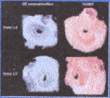
Taken from three-dimensional echocardiographic and stereolithographic replicas, the next illustration demonstrates mitral valve stenosis (marked narrowing of the mitral valve orifice or opening down to an area of 1 to 2cm2), analogous to a funnel with the mouth being the entire circumference of the mitral valve ring and the narrowed stem or spout (opening) being similar to the narrowed opening of the mitral valve, as a result of scar tissue fusing the leaflets of the valve together to impede blood flow into the left ventricle and to reduce mitral valve motion (see figures 44A-D) (as can occur in rheumatic fever, see definition). The first upper picture on the left is looking from the left atrium at the mitral valve in diastole (phase of blood flow from left atrium into the left ventricle). The bottom left picture of the mitral valve is looking from the left ventricle. Note the doming of mitral valve due to the fusion and scarring deformity. Also, the black centers in the mitral valve illustrations represent the size and shape of the mitral orifices. The pictures on the right are stereo lithographic replicas of the above mitral valve. The reduced mitral valve orifice causes an increase blood flow velocity through the valve into the left ventricle to deliver the normal volume of oxygenated blood to the body. There is also an increase pressure in the left atrium due to the reduced opening of the valve (see figure 44E). In time the pressure can increase to the point where it is transmitted back to the lungs resulting in congestion, fluid accumulation (edema) in the lungs and shortness of breath and reduced exercise tolerance.
Reference:Binder,T.M.,Moertl,D.,Mundigler,G.,Rehak,G.,Frankee,M.,Delle-Karth,G.,Mohl,W.,Baumgartner,H.,Mauer,G., JACC,vol.35,Jan. pages230-237,modified