Figure 35b
Two-dimensional
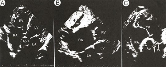
subcostal views from a patient with chronic obstructive lung disease.
The liver (L) is seen anteriorly in each frame.
A.
Five-chamber view, directed to include a portion of the inferiror vena
cava (IVC) at its junction with the right atrium (RA), shows the aorta
(Ao) between the cardiac septa and atrioventricular valves in addition
to the right and left (LA) atria and right (RV) and left (LV) ventricles.
The cardiac apex is not seen in this view.
B.
Enlargement of the four-chamber view to best show the junction of the
inferior vena cava (IVC) and the right atrium. The hepatic vein (HV)
empties into the inferior vena cava. Complete imaging of the interatrial
septum is appreciated in this view.
C.
Diastolic frame of the short-axis right ventricular outflow tract plane.
The aorta (Ao) occupies the central portion of the image. Two of the
open leaflets of the tricuspid valve (TV) and the closed pulmonic valve
(PV) are identified.
J.M. Felner, R.P. Martin, The Echocardiogram, The Hurst's The Heart, 8th ed., p 391.