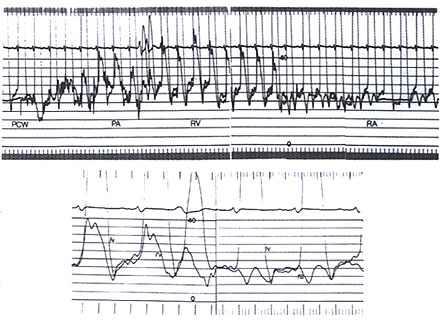
Figure 38 h
Hemodynamic record of a patient with surgically proven constrictive pericarditis. Top. Slow paper speed recording of high-gain left ventricular (LV) pressure and simultaneous right heart pullback from pulmonary capiLlary wedge (PCW) to pulmonary artery (PA), right ventricle (RV), and right atrium (RA). Bottom. Fast paper speed recording of LV and simultaneous RV and RA pressure tracings. Note the increased and equal atriaL and diastolic pressures, the prominent X and V descents on the RA tracing, and the dip and plateau on the RV and LV tracings during longer diastotes.
(Courtesy of Peter j. Engel, MD. From
Hoit BD. Pericardial disease and pericardial
heart disease. In: O'Rourke RA, ed. Stein's Internal Medicine, 5th ed.
St. Louis, Missouri: Mosby-Vear Book; 1998:273.)