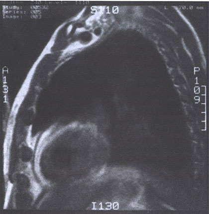
Figure 38 j
MRI scan (spin-echo image) from a patient
with constrictive pericarditis. The pericardium is viewed as a Line of
low signal intensity (black) sandwiched between higher-intensity epicardial
and pericardial fat (white). Note the regional variation of pericardial
thickness, which is normally 1 to 2 mm.
(From Hoit BD. Pericardial disease and pericardial heart disease. In: ORourke RA ed. Stein's Internal Medicine, 5th ed. St. Louis, Missouri: MosbyYear Book;1998:273.