Figure
39a
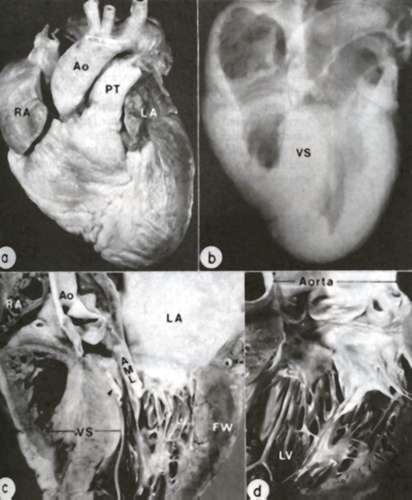
Anatomic
features of HCM are demonstrated in the heart of a 26-year-old man.
A. Exterior view; both right atrium
(RA) and left atrium (LA) are dilated. Ao= aorta; PT= pulmonary trunk.
B. Radiograph of specimen showing
asymmetric thickening of ventricular septum (VS).
C. Coronal section ; the septum
is clearly thicker than left ventricular free wall (FW); an endocardial
mural contact plaque (arrow) is present in the left ventricular outflow
tract in apposition to the anterior mitral leaflet (AML).
D. closer view of plaque and thickened
anterior leaflet.
(From WC Roberts, VJ Ferrans: Hum Pathol 6:287-342, 1975.)