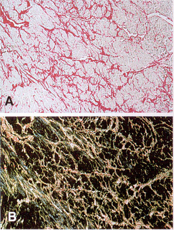
Figure 39e
Transversely cut cardiac muscle cells in ventricular septum from a 17-year-old patient with HCM, stained with picrosirius red and viewed by light (A) and polarization (B) microscopy. The myoctes show characteristic encasement within a dense network of matrix collagen. (Magnification x100).