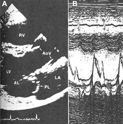
Figure 45
Echocardiogram from a patient with mitral valve prolapse. A. Parasternal long-axis view in systole show the posterior leaflet (PL) of the mitral valve with superior displacement (bowing) into the left atrium (LA), i.e., arching posteriorly above the level of the atrioventricular groove and behind the normal coaptation point of the left atrium. B. M-mode study shows abrupt posterior displacement in midsystole (arrow) of the posterior mitral leaflet. There are multiple echoes arising in the area of the valve during systole as a result of the redundancy of the leaflets. These findings are characteristic of mitral valve proplapse. LV, left ventricle; AL, anterior leaflet of mitral valve; RV, right ventricle; AoV, aortic valve.
J.M. Felner, M.D., R.P. Martin, M.D., The Echocardiogram, The Hurst's The Heart, 8th ed., p 405.