Figure 48g
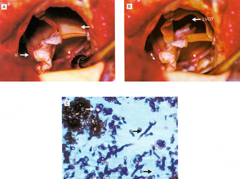
A 77-year old man presented with a two month history of fever, chills, and malaise. Three months earlier, he had undergone valve replacement for calcific aortic stenosis at another instituition. Multiple subsequent blood cultures were positive for Candida albicans. Clinica l signs of endocarditis (new murmurs, splenomegaly, Osler's nodes, and retinal lesions) were absent.Transesophageal echocardiography identified a mobile vegetation within the leaflets of the prosthetic valve that prolapsed into the ventricle during diastole.The patient as treated with amphericinB and flucytosine for two weeks. At operation the prosthetic valve (PanelA) contained a whitish vegetation(V) attached to the leaflets(L).The vegetation extended into the left ventricular outflow tract (LVOT) when a leaflet was opened (Panel B). A photomicrograph of the vegetation demonstrated innumerable candidal element, including pseudomyceli(P), blastospores(B),and chlamyspores(C);(Panel C; silver stain, x40).The prosthetic valve was removed, and a aortic-valve homograph was implanted(i.e., a mini-root replacement) to reconstruct the heart. Postoperatively, the patient received liposomal amphtericinB and flucytosine followed by oral fluconazole, and the sepsis cleared.
Reference:Tuna,I. And Harrison,M.,N.England Journal Med.,Vol.344,No.4,Jan.25,2001,Pp.275