Figure 51f
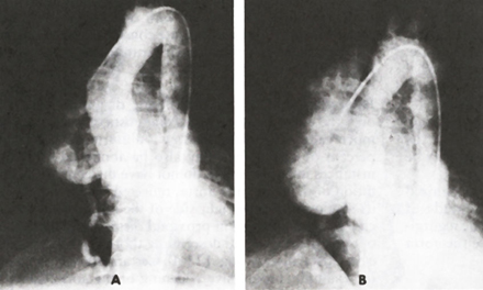
Serial frames from an aortogram of a patient with type I aortic dissection. Contrast material is seen to fill a tremendously dilated false channel. This accounts for the dilatation of the ascending aorta noted in figure 51E.
|
|
|
Figure 51f Serial frames from an aortogram of a patient with type I aortic dissection. Contrast material is seen to fill a tremendously dilated false channel. This accounts for the dilatation of the ascending aorta noted in figure 51E. |