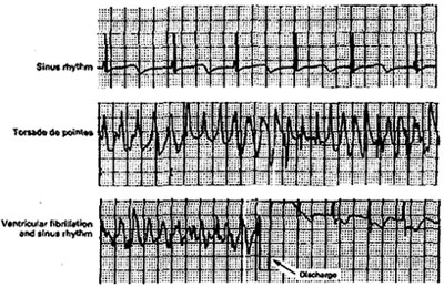
Figure 62
Internal Ventricular Defibrillation.
A 12-year-old boy with the familial long-QT syndrome had an automatic
cardioverter-defibrillator implanted after resuscitation from cardiac
arrest. The device was programmed to wait 7.5 seconds before discharging
in response to ventricular tachycardia or ventricular fibrillation.
The boy took 120 mg of nadolol daily. Three months later, he suddenly
collapsed while answering a question in school. Analysis of the cardioverter-defibrillator
revealed the following sequence of events: normal sinus rhythm with
a corrected QT interval of 0.66 second (top tracing ) was followed by
torsade de pointes (middle tracing) and ventricular fibrillation (bottom
tracing), prompting discharge of the cardioverter-defibrillator (arrow)
and a return to sinus rhythm within 600 msec after discharge. (The rhythm
strips are not continuous.) The boy regained full consciousness within
two minutes, with no residual neurologic deficits. During the next 15
months, he had no syncope or evidence of recurrent ventricular tachyarrhythmias.
Genotypic analysis revealed a mutation in the HERG gene, a gene encoding
a potassium channel.
Moss, A.J., M.D., Daubert, J.P., M.D., Internal Ventricular Defibrillation, The New England Journal of Medicine, Feb 10, 2000, p 398.