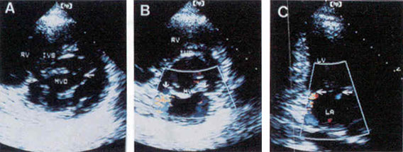
Figure 69
Both commissural splitting and commissural mitral regurgitation after successful PMV (Percutaneous Mitral Valvuloplasty) on parasternal short axis view (A and B) and apical two-chamber view (C). The arrows indicate split commissures (A) and origination of MR from both commissures (B and C). IVS, interventricular septum; LA, left atrium; LV, left venricle; MV, mitral valve; MVO, mitral valve orifice; RV, right ventricle. PMV involves the insertion of a balloon into the stenotic mitral valve orifice to accomplish dilatation.
Kang, D.H., MD, Park, S.W., MD, Song, J.K., MD, Kim, H.S., MD, Hong, M.K., MD, Kim, J.J., MD, Park, S.J., MD, Long-Term Clinical and Echocardiographic Outcome of Percutaneous Mitral Valvuloplasty, Journal of the American College of Cardiology, Vol. 35, Jan 2000, p 169-175. (modified)