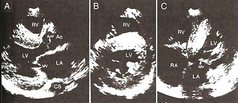
Figure 73a
Parasternal Long- and Short-Axis Views
of the Left Ventricle, Showing Symmmetric Left Ventricular Hypertrophy.
Small regions of increased reflectivity are apparent within the interventricular
septum (long arrows). The aortic-valve cusps are thickened and also
show increased reflectivity (short arrow). This is an illustration of
how the echocardiogram can pick up abnormalities as above in the case
of amyloidosis of the heart.
DiSalvo, T.G., King, M.E., Smith,
R.N., Case Records of the Massachusetts General Hospital, A 66-Year-Old
Woman with Diabetes, Coronary Disease, Orthostatic Hypotension, and
the Nephrotic Syndrome, NEJM, Vol. 342, Jan 27-00, p 264-274.(modified)