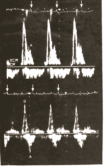
Figure 74a
Pulsed Doppler Flow Patterns of Transmitral Inflow (Upper Panel) and Pulmonary Venous Inflow (Lower Panel), with Characteristic Features of Restrictive Cardiomyopathy (in this case due to amyloid deposition). The upper panel shows a prominent early diastolic filling wave (E) with rapid deceleration and a smaller atrial filling wave (A). The lower panel shows blunted pulmonary venous inflow in systole (S), with a prominent diastolic filling wave (D), which decelerates rapidly, followed by a reversal of flow into the pulmonary veins in late diastole during atrial contraction (A). The arrows in both panels show the R wave of the electrocardiogram (EKG). This illustration demonstrates the use of the Doppler echocardiogram to study blood flows in and out of various chambers of the heart to make diagnoses.
DiSalvo, T.G., King, M.E., Smith, R.N., Case Records of the Massachusetts General Hospital, A 66-Year-Old Woman with Diabetes, Coronary Disease, Orthostatic Hypotension, and the Nephrotic Syndrome, NEJM, Vol. 342, Jan 27-00, p 264-274. (modified)