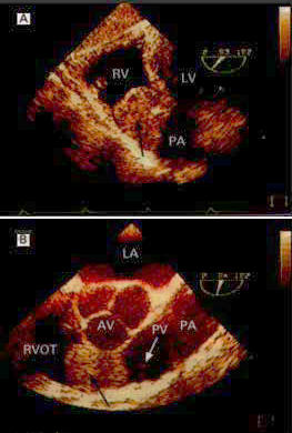
Figure 80
Transesophageal Echocardiograms of a Right Ventricular Myxoma. Panel A shows a transgastric view of the myxoma (arrow) in the dilated right ventricle. Panel B shows a midseophageal view of the myxoma (black arrow) occluding the right ventricular outflow tract (RVOT). RV denotes right ventricle, LV left ventricle, PA pulmonary artery. This study is from a 74 year old man with symptoms of shortness of breath, fainting, and dizziness due to the obstruction of blood flow through the right ventricular outflow tract and pulmonary valve from an uncommon cardiac myxoma arising from the right side of the IVS.