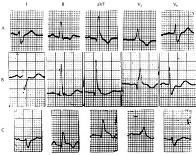
Figure 94-36
Left posterior fascicular block with
RBBB.
A. No MI.
B. Anteroseptal MI (note Q wave in V2).
C. Inferior MI (note ST-segment elevation and T-wave inversion in leads
11 and AVF with slight ST-segment depression in lead in lead 1). The
difference between A and C are not very marked since pure LPFB may produce
an almost abnormal Q wave in the inferior leads. Conversely, this conduction
disturbance, by producing an initial depolarization wave front oriented
superiorly, is capable of decreasing the depth(thus normalizing) the
abnormal Q-wave characteristic of inferior MI in leads11, 111, and AVF.