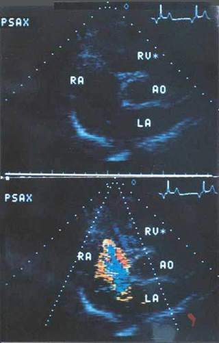
Figure 112b
Ostium
secundum atrial septal defect, visualized by color Doppler flow imaging
in a low parasternal short-axis orientation.
Top. Anatomic image demonstrating the aorta,
left atrium, right atrium, and right ventricle. Although the chambers
of the right side of the heart are large, dropout of echoes in the interatrial
septum in this view may occur because the atrial septum and the ultrasound
beam are nearly parallel.
Bottom. The color flow jet shows a central
aliased portion (coded blue) surrounded by unaliased flow (coded red-orange).
This clearly proves the existence of a large secundum defect with left-right
shunting. PSAX, parasternal short axis; Ao, aorta; LA, left atrium;
RA, right atrium; RV, right ventricle.
Hurst's The Heart, 8th edition, Plate XI, 1994.