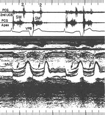
Figure 117
Phonocardiogram
and echocardiogram in mitral valve prolapse.
A. The phonocardiogram shows a high-frequency holoysystolic murmur (SM)
with late systolic accentuation due to regurgitation of blood through
the mitral valve into the atrium. A low-frequency mid-diastolic murmur
(MDM) is present at the apex.
B. The echocardiogram demonstrates a hammock-shaped systolic motion
of the valve leaflets. The rhythm is atrial fibrillation with bigeminy.
1= first heart sound; 2= second heart sound; MVE= mitral valve echogram.
(Courtesy of Dr. Ernest Craige.)
Gaasch, W.H., M.D., O'Rourke,
R.A., M.D., Cohn, L.H., M.D., Rackley, C.E., M.D., Mitral Valve Disease,
Hurst's The Heart, 8th edition, 1994, p 1507.
(modified)