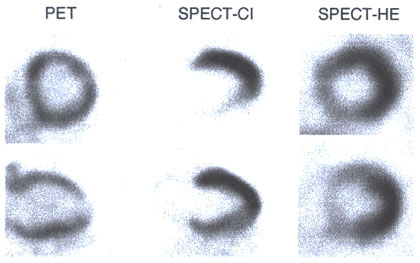
Figure 216-1
Examples of myocardial [18F] deoxyglucose images obtained with PET (left), a SPECT-like system with coincidence detection (CI, middle panel) (courtesy Dr. R. Henkins, Chicago, IL), and a SPECT system equipped with an ultra-high-energy-photon general-purpose collimator (HE). For the SPECT-CI images, the non-attenuation-corrected image is shown on top and the corrected image at the bottom.