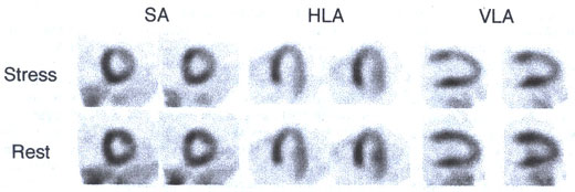
Figure 216-4
Stress induced perfusion defect in the lateral wall of the left ventricle, depicted each on two contiguous short- (SA), horizontal long- (HLA), and vertical long-axis slices (VLA) through the mid-left ventricle. Note the normalization of myocardial perfusion on the rest images.