Figure 35a
Family
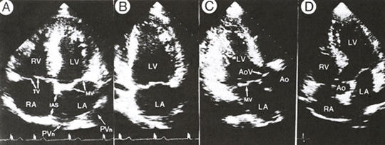
of two-dimensional apical views from a normal subject.
A. Systolic frame of the four-chamber view
shows the relations of the cardiac chambers, atrioventricular valves,
and the interatrial and interventricular septa to each other . Note
that the insertion of the tricuspid valve (TV) is inferior (mor apical
) to that of the mitral valve (MV) and that the common problem of partial
drop-out of the midportion of the interatrial septum (IAS) is apparent.
The entry of the pulmonary veins (Pvn) into the left atrium (LA) is
clearly seen.
B. Systolic frame of the two-chamber view shows the left ventricle and
left atrium with the closed mitral valve.
C. Systolic frame of the long-axis view shows the aortic valve (AoV)
and the proximal portion of the ascending aorta (Ao). The anterolateral
and postermedial walls of the left ventricle (LV) and the left atrium
are also seen. The posteromedial papillary muscle, arising near the
apex, may be seen in this view. D. Five-chamber view includes a portion
of the aorta (Ao) seen at the junction formed by the cardiac septa and
atrioventricular valves as well as the right (RA) and left atria and
right (RV) and left ventricles.
J.M. Felner, R.P. Martin, The Echocardiogram, The Hurst's The Heart, 8th ed., p 390.