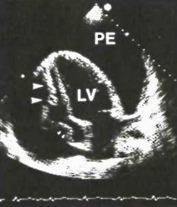
Figure 36a
Two-dimensional apical four-chamber view from a patient with cardiac tamponade. This diastolic frame shows a large pericardial effusion (PE) that completely surrounds the heart. The right ventricular cavity is virtually nonexistent. There is both right ventricular wall collapse (large arrows) and right atrial wall collapse (small arrows) consistent with tamponade. LV, left ventricle.
J.M. Felner, R.P. Martin, The Echocardiogram, The Hurst's The Heart, 8th ed., p 402.