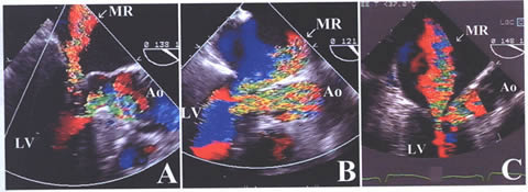
Figure 39j_b
Transesophageal echocardiography images
in three patients with hypertrophic obstructive cardiomyopathy showing:
(A) Posteriorly directed mitral regurgitation
jet (arrow) in a patient without independent abnormality of the mitral
valve.
(B) Anteriorly directed mitral regurgitation
jet (arrow) in a patient with posterior mitral valve leaflet prolapse.
(C) Centrally directed jet of mitral
regurgitation (arrow) in a patient with mitral stenosis.
Ao = aorta; LV = left ventride; MR mitral regurgitation.