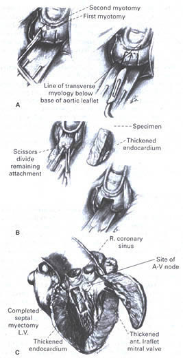
Figure 39l
Illustration of ventrucutar septal
myotomy-myectomy operation (Morrow procedure).
A. Two verticaL parallel myotomies are made in the cephalad portion
of the septum about 1 cm apart. Transverse incision is then made, connecting
the two parallel myotomies.
B. Attachments of the muscle bar to the septum are divided; this segment
of muscle is isolated and then excised.
C. After completion of the myotomy-myectomy, a rectangular channel about
4 cm long and 2 cm wide is evident extending from the aortic annulus
to a point just distal to the caudal margins of the mitral leaflets.
(From Maron BJ et al,with permission from the authors and the European Heart Journal.)