Figure 41
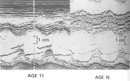
Development and progression of left ventricular
hypertrophy in children with HCM. Upper panel:
Development of marked hypertrophy of the anterior basal ventricular
septum (VS). M-mode echocardiograms shown here were obtained at the
same cross-sectional level in a girl with a family history of HCM. At
age 11, ventricular septal thickness was at upper limit of normal (10
mm); at age 15, septal thickness had increased markedly (to 33 mm),
and appearance of the echocardiogram is typical of JCM. The patient
remained asymptomatic throughout this period of time but died suddenly
and unexpectedly at age 17. PW= posterior left ventricular free wall.
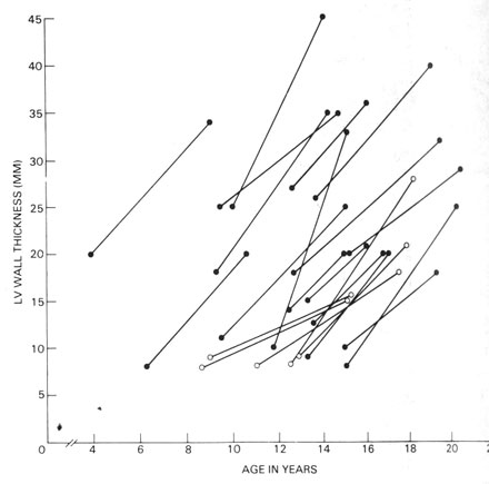
Lower panel: Dynamic, striking changes
in left ventricular wall thickness with age in 22 children, each patient
is represented by the left ventricular segment that showed the greatest
change in wall thickness. Open symbols denote five patients who had
no evidence of hypertrophy in any segment of the left ventricle at the
initial evaluation but subsequently developed de novo hypertrophy typical
of HCM.
(From BJ Maron et al: N Engl J Med 315:610, 1986)