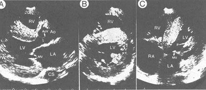
Figure 43a
Two-dimensional echocardiogram from
a patient with confirmed amyloid heart disease.
A. Parasternal long-axis view in diastole shows right ventricular hypertrophy,
concentric left ventricular hypertrophy, and bright reflectors of the
interventricular septum (VS), characteristic of amyloid involvement
of the myocardium. The left ventricular (LV) cavity is normal in size,
but the left atrium is dilated. The mitral valve (MV) appears thickened.
B. Short-axis view at the level of they hypertrophied papillary muscles
(P) also shows right ventricular hypertrophy, concentric left ventricular
hypertrophy, and a general increase in refractile pattern.
C. Apical four-chamber view again shows the general increase in the
refractile pattern of the septum and the hypertrophied papillary muscle
(P) and chordae tendineae. The fact that the interatrial septum does
not have significant dropout suggests that it, too, is hypertrophied.
RV, right ventricle; CS, coronary sinus; PW, posterior wall; AoV, aortic
valve; Ao, aorta ; RA, right atrium.
J.M. Felner, M.D., R.P. Martin, M.D., The Echocardiogram, The Hurst's The Heart, 8th ed., p 403.