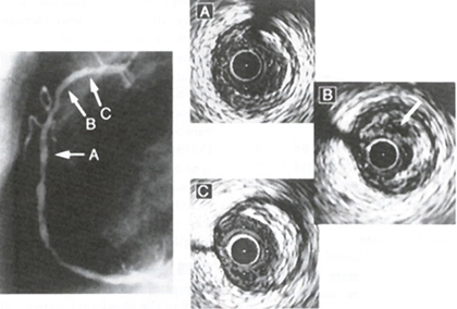
Figure 66
Representative intravascular ultrasound image of vulnerable atherosclerotic plaque (see fig 70). (Left) The right coronary angiography showed multiple luminal irregularities. (Right) (A) At the distal portion, there existed mild concentric lesion. (B) In the proximal portion there was a significant eccentric lesion in which the echolucent area with percent plaque area of 67% was seen (arrow). (C) At the very proximal of this artery, there was also eccentric lesion with relatively high echo density. All three lesions were examined at follow-up.
Yamagishi, M., MD, Terashima, M., MD, Awano, K., MD, Kijima, M., MD, Nakatani, S., MD, Daikoku, S., MD, Ito, K., MD, Yasumura, Y., MD, Miyatake, K., MD, Morphology of Vulnerable Coronay Plaque: Insights from Follow-up of Patients Examined by Intravascular Ultrasound Before an Acute Coronary Syndrome, Journal of the American College of Cardiology, Vol. 35, Jan 2000, p 106-111. (modified)