Figure 77e
One
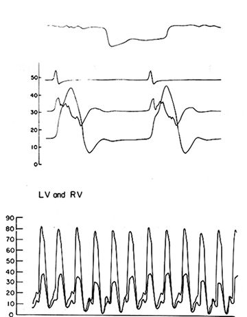
right ventricular pressure tracing typical of restrictive amyloid cardiomyopathy
showing ( top ) the characteristic dip and and
plateau configuration.
But in the bottom part showing both right and left ventricular tracings
superimposed the plateau is absent despite the diagnosis.