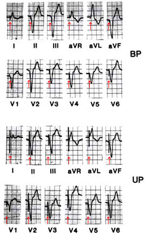
Figure 85
Twelve-lead ECGs demonstrating the differences between bipolar (BP) and unipolar (UP) pacing. The illustrations are from the same patient with endocardial pacing from the apex of the right ventricle. In BP pacing (top), small stimulus artifacts are seen except over the anterolateral leads (V3 to V6), which physically lie closest to the lead poles. In UP pacing (bottom), stimulus artifacts are prominent in all leads. I both ECGs, there is a left bundle branch block appearance with no R wave in the lateral chest leads. The frontal plane axis is to the extreme left.
Mond, H.G., MD, Permanent Cardiac Pacemakers: Techniques of Implantation, Testing, and Surveillance, Hurst's The Heart, 8th ed., p 815-841