Figure 86
Electrocardiogram
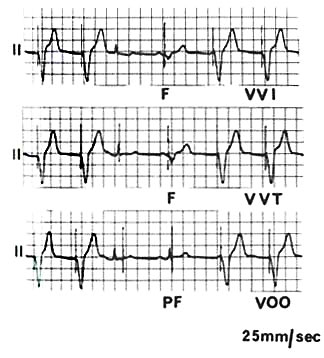
lead II demonstrating three modes of ventricular pacing. In each example
the first two and the last two beats are paced. The third and fourth beats
are sinus beats.
Top: VVI pacing. The third beat is sensed with inhibition of ventricular
output (meaning that the pacemaker did not fire to excite the ventricle
itself). The fourth beat is a fusion beat (F, meaning that the normal
sinus beat and the pacemaker beat fused).
Middle: VVT pacing. The third beat is sensed and the pulse generator
discharges into the latter part of the QRS but, unlike a fusion beat,
does not contribute to the wave of depolarization. Depending on the timing
of QRS detection, VVT pacing may deform the QRS complex. The fourth beat
is a true fusion beat.
Bottom: VOO pacing. There is no QRS sensing and the stimulus artifact
falls after the QRS in the refractory period (so the pacemaker failed).
With the fourth beat the stimulus artifact is close to the QRS and is
therefore a form of pseudofusion (PF). (see fig 88 for explanation of
abbreviations)
Mond, H.G., MD, Permanent Cardiac Pacemakers: Techniques of Implantation, Testing, and Surveillance, Hurst's The Heart, 8th ed., p 815-841