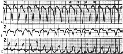
Figure 9a
A. VT with regular independent P waves (arrows) B. VT with retrograde conduction to the atria (arrows for P waves) C. VT with fusion beats (arrows) with sinus P waves before each fusion beat.
Myerburg, R.J., M.D., Castellanos, A., M.D., Kessler, K.M., M.D., Recognition, Clinical Assessment and Management of Arrhythmias and Conduction Disturbances, Hurst's The Heart, 8th ed, p 738.