narrowing
of aortic valve orifice
In patients younger than 65 years old with
symptomatic aortic valve stenosis, the most frequent pathologic
finding is a bicuspid aortic valve (normally tricuspid), which
occurs in 2-3% of the population, being four times as common
in men and boys than in women and girls.
20% of cases with bicuspid valve have an
associated heart abnormality, like a patent ductus arteriosus
or aortic coarctation.
The bicusp valve has a simple fused commissure
and eccentrically oriented orifice ( see figures 46a,
46b,
46c,
47
).
Due to the stresses of pressure, the valve
becomes thickened and calcified, leading to reduced motion.
Calcific valvular disease represents the end stage of stage
of an active disease process. In the early stages, the aortic
side of the valve contains focal lesions characterized by thickening
of the subendothelium and the adjacent fibrosa. These lesions
contain low density-lipoproteins, Lp(a) lipoproteins, macrophages,
and T lymphocytes. Areas of microscopic calcification form within
regions of lipoprotein accumulation, and some macrophages within
lesions produce osteopontin, a protein that modulates tissue
calcification.
This stage of the disease process is evident
on echocardiography as a mild, irregular leaflet thickening
without obstruction of ventricular outflow and is termed aortic
sclerosis. As the disease progresses, calcification and fibrosis
increase leaflet stiffness and reduce systolic opening, eventually
leading to a reduction in the area of the valve and an increase
in forward velocity.
Clinically significant obstruction of flow
through the valve is present in about 1 to 2 % of adults over
the age of 65 years, and it is likely that most of these patients
will ultimately have symptoms necessitating valve replacement.
Obstruction of left ventricular outflow results
in pressure overload, with compensatory hypertrophy of the left
ventricle to maintain normal wall stress, preserving systolic
function, which can improve after valve replacement. Clinical
outcome is most closely related to the presence or absence of
symptoms.
Once symptoms occur, the clinical outcome
is extremely poor, with two year survival rates below 50%. It
is well established that this dismal prognosis can be reversed
by valve replacement with acceptable levels of operative mortality
and morbidity and postoperative survival similar to that of
age-matched normal adults.
In symptomatic adults with systolic murmurs,echocardiography
is essential to identify those likely to benefit from surgery.
Surgery should be considered even for elderly persons and those
with left ventricular dysfunction, since they often improve
with valva replacement.
But adults without symptoms have an excellent
prognosis.The simplest measure of the extent of stenosis is
the forward velocity across the aortic valve. This velocity
is about 1.0 m per sec. in normals and increases to 2.5 to 2.9
m per sec. in mild stenosis,3.0 to 4.0 m per sec. in moderate
stenosis, and more than 4.0 m per sec. in severe stenosis.
Measurement of the area of the valve is useful,
being above 1.5 cm2 in mild disease, 1.0 to 1.5 cm2 in moderarte,
and in severe disease less than 1.0 cm2.
There is a substantial variation in the degree
of stenosis associated with the onset of symptoms; as a result,
many asymptomatic patients with severe obstruction are now identified
by echocardiography. Although some feel that valve replacement
should be preformed in patients with severe aortic stenosis
before the onset of symptoms, others feel that the optimal time
for surgery is when symptoms develop. Periodic echocardiography
is appropriate to determine when to operate. Once symptoms supervene,
prompt valve replacement surgery is indicated.
Reference:Otto,C.M.,N Engl J Med.Vol.343,No.9,652-654.
Reference:Rosenhek,R. AND OTHERS,N Engl J MED.,vOL.343,N.9,611-624.
Dilatation of the aortic root may occur as
a sequelae of the stenosis.
The normal adult aortic valve opening is
3.0-4.0cm2. Aortic stenosis becomes hemodynamically significant
when the area is about 1cm2 to 0.8cm2 (as noted above in the
above discussion of the area of the valve orifice which can
be classified as to severity of stenosis), as the systolic flow
is impeded across the valve.
Diagnosis is suggested by the detection of
an ejection high pitched click, a harsh diamond-shaped crescendo,
decrescendo, basal systolic heart murmur transmitted into the
neck, a soft or absent aortic component of the second heart
sound by auscultation, a palpable apical thrust reflecting enlargement
of the left side of the heart confirmed by electrocardiography
(increased amplitude of the QRS complex with ST and T wave changes,
increased amplitude of S waves in the right precordial leads,
increased amplitude of the R wave in the left precordial leads,
depression of the ST segment and inversion of the the T wave,
(see figure
24 )
Doppler echocardiography and echocardiography
can determine the severity of the stenosis including degree
of thickening, calcification,and reduced mobility of the aortic
leaflets and assess the left ventricle contraction function
and wall thickness regarding the presence of left ventricular
hypertrophy. A bicuspid valve aortic valve can be recognized
by the asymmetry of the two leaflets ( figure
47 ).
The doppler tecnique allows measurement of
the velocity across the aortic valve, which provides good estimates
of the systolic gradient ( figure 46f
), and using the continuity equation the aortic valve area can
be calculated ( figure
46e ).
Heart catherterization can be used to judge
severity of the valve stenosis (if undeterminable non-invasively)
by recording the gradient across the valve, estimating the stenotic
area ( figure
46d ), evaluating the left ventricular function and to determine
if coronary artery disease is concurrently present.
Symptoms include chest pain (exertional, angina
pectoris), fainting, and heart failure (exertional shortness
of breath, nocturnal cough, orthopnea, paroysmal nocturnal dyspnea,
hemoptysis). The syncope may related to decreased left ventricular
cardiac output due to myocardial ischemia both during and separate
from exertion or to cardiac arrhythmias (which may also account
for episodes of sudden death).
Adults with asymptomatic stenosis have a
normal life expectancy, but should receive antibiotic prevention
against infection of the aortic valve(see above discussion).
If symptoms develop, survival is limited.
Hence, valve replacement should be performed promptly. In asymptomatic
younger individuals, however, the documentation of severe aortic
stenosis is, in itself, an indication for intervention. Mild
aortic stenosis in asymptomatic patients with gradients below
50mm warrants careful surveilance.The management of patients
in the intermediate group (gradients 50-75mm Hg) is more controversal,
but evidence argues in favor of elective intervention.
Surgery in the young adult with congenital
aortic stenosis must be considered as palliative. In the absence
of calcification, aortic valvotomy is the procedure of choice.
Perioperative mortality in adolescents and adults is extremely
low and late survival is excellent. As surgical valvotomy is
palliative, catheter balloon valvotomy has obvious attractions
as the initial procedure or as treatment for restenosis.
Valve replacement is the only option for valves
unsuitable for valvotomy, including those with significant calcification
and regurgitation.
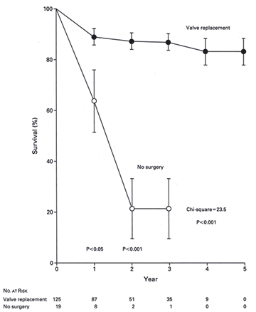
Late Results of Aortic Valve Replacement
Late morbidity and mortality in patients after
aortic valve replacement are generally quite satisfactory. Actuarial
survival at 5 years with various types valve prostheses is about
80% (See Figure 203 Immediately Below).
The degree of left ventricular dysfunction,
as well as concomitant coronary disease and coexiting morbid
conditions are significant determinants of late mortality. Late
deaths are most often due to chronic congestive heart failure,
thromboembolic stroke, myocardial infarction in those with coronary
disease, and cardiac arrhythmias leading to sudden death, erspecially
in those with an enlarged dilated heart.
Sudden death after aortic valve replacement
is most commonly thought to due to ventricular arrhythmias,
but it may also result from thromboemboli. The risk of thromboembolism
is much less after aortic valve replacement than after mitral
valve replacement because most patients are in sinus rhythm
( figure 180 ). The thromboembolic risk is lowest after a homograft
or pulmonary autograft valve replacement, following which it
is virtually zero. Patientswith porcine or pericardial valves
have a risk of about 1.5 events per 100 patient years, and in
patients with prosthetic valves the risk is 1.5 and 2.5 events
per 100 patient years. Thrombosis of prosthetic valves was a
more frequent complication with previous prosthetic valves,
but it now occurs rarely and only when anticoagulation is stopped
for a long period of time. With newer, low-profile, more hemodynamically
efficient valves, this morbidity has decreased, and warfarin
anticoagulation provides excellent protection from thromboembolism
for most patients. It is recommended that patients with mechanical
prosthetic valves be maintained on levels of wararin that prolong
the INR to 2.5 to 3.5.
Patients with biologic aortic valves, of course,
have the risk of late structural valve degeneration. The probablity
of freedom from valve reoperation at 10 years in patients witha
cryopreserved homograft is about 90%; this is somewhat better
than the porcine bioprostheses, where the freedom from structural
valve degeneration is about 90% at 8 years and 40% at 15 years.
In the aortic position, a bioprosthetic valve is the valve of
choice in most elderly patients because the rate of valve tissue
degeneration is considerably lower than in younger age groups
and the probability of valve tissue failure in patients over
70 is 20% at 10 years. The use of tissue valves in the elderly
obviates the use of anticoagulation, which is very advantageous.
In the younger age group, mechanical prosthetic valves tend
to be used because most patients wish to minimize the probability
of reoperation due to valve dysfunction and because of the accelerated
fibrosis-calcification in biologic prosthetic valves in children
and young adults. An exception is the child-bearing-age female,
who should not anticoagulation during pregnancy because of the
teratogenic potential of warfarin in the first trimester, and
who should receive a biologic valve if replacement is indicated.
Reference:Rapaport,E. and Others,Aortic Valve
Disease,Hurst's,Heart,8th Edition,PP1457-1466
It is debatable whether or not any patient
who has had significant obstruction should be allowed to participate
in competitive sports. A residual gradient greater than 20 mmHg
or persistent left ventricular hypertrophy are considered to
be contraindications to vigorous physical activity.
Reference:Deanfield,J.E.
and others,Adult Congental Heart Disease,Hurst's THE HEART,8TH
edition, 1829-1853.
Pathophysiology
Of Aortic Stenosis
The AVA has to be reduced by about 50%
of normal before a measurable
gradient can be demonstrated in humans.In most patients with
AS ,cardiac
output is in the normal range and initially increases normally
with
exercise. Later, as the severity of AS increases progessively,the
cardiac
output remains within the normal range at rest,but,on exercise,
it no
longer increases in proportion to the amount of exercise undertaken
or
does not increase at all (fixed cardiac output). With the development
of
heart failure,there is a reduction in the resting cardiac output
and a
tachycardia.As a result,stroke volume may be so lowered that
it results
in a small gradient across the left ventricular outflow tract
in spite
of severe AS.
Echo/Doppler, when properly
applied , is extremely useful for estimating
the valve gradient and AVA noninvasively.When compared with
results
obtained at cardiac catheterization,the standard error of the
mean
gradient in the best laboratories is 10mmHg.Thus,the mean gradient
by
Doppler can be expected to be within +- 20mmHg (95% confidence
level) of
that obtained at catheterization.
Table
1 Suggested Conservative Guidelines for Relating Severity of
Aortic Stenosis to Doppler Gradients
in Adults with Normal Cardiac Output and Normal Average Heart
Rate
Peak
Gradient---------- MeanGradient
--------------------------------------------------------Severe
AS
mmHg ---------------------mmHg
Approx.80------------------70
--------------------------Highly likely
60-79----------------------50-69-----------------------
Probable
<60-----------------------
<50---------------------------Uncertain
Rahimtoola,S.H.,Aortic Valve Disease,Hurst's Diseases of The
Heart,10th Edition,Vol.2,Pp.1682-1695.
Cardiac
cathetrization remains the standard technique to assess the
severity of AS "accurately".
As
indicated in the table below, AS can be considered severe when
the valve area is 1.0 cm2 or less or the AVA index is 0.6 per
square meter or less.
TABLE
2 A Suggested Grading of the Degree of Aortic Stenosis
Aortic Stenosis , AVA, cm2 , AVA Index, cm2/m2
Mild------------ >1.5 ,------------->0.9
Moderate-------->1.0-1.5,
-------->0.6-0.9
Severe------------0.8-1.0
,--------- 0.4--0.6
Rahimtoola,S.H.,Aortic
Valve Disease,Hurst's Diseases of The Heart,10th Edition,Vol.2,Pp.1682-1695.
Natural History and Prognosis
Valvular
AS is frequently a progressive disease, the severity increasing
over time. The factors that control this progression and the
time it takes for severe outflow obstruction to develop are
unknown; however, it appears that in the older patient, AS may
progress at about twice the rate that it does in the younger
patient. In a study of 142 patients with "mild" stenosis
(catheterization-proven AVA >1.5 cm2), the rate of progression
to severe stenosis was 8 percent in 10 years, 22 percent in
20 years, and 38 percent in 25 years. At 25 years, 3 percent
still had mild AS. The duration of the asymptomatic period after
the development of severe AS is also unknown; some recent data
suggest that it maybe less than 2 years. The outcome of the
asymptomatic patient with severe AS is not known. In the study
of 123 asymptomatic patients aged 63 ± 16 years, the
actuarial probability of death or aortic valve surgery was 7
± 5 percent at 1 year, 38 ± 8 percent at 3 years,
and 74 ± 10 percent at 5 years. The event rate at 2 years
for peak aortic jet velocity by Doppler ultrasound of >4
m/s was 79 ± 18 percent, for 3 to 4 m/s was 66 ±
13 percent and for <3 m/s was 16 ± 16 percent. However,
the limitations of gradients and of aortic peak velocity obtained
by Doppler ultrasound should be kept in mind. The overwhelming
majority of adults with severe AS who are seen by cardiologists
have symptoms.
Severe disease in adults is lethal, particularly if the patient
is symptomatic, with a prognosis that is worse than for most
forms of neoplastic disease. The 3-year mortality is approximately
36 to 52 percent, the 5-year mortality is about 52 to percent,
and the 10-year mortality is 80 to 90 percent. A recent study
of elderly patients (average age 77 years) showed 1-year and
3-year mortalities were 44 and 75 percent, respectively. With
the onset of severe symptoms (angina, syncope, or heart failure),the
average life expectancy is 2 to 3 years.Almost all patients
with heart failure are dead in 1 to 2 years.A combination of
symptoms is much more ominous, a sign of a greatly reduced survival.Sudden
death, like syncope, occurs in the presence of severe AS.Its
wexact incidence is difficult to determine but may be about
5%.
TABLE 3 Natural History of Mild Aortic Stenosis (n=142), AVA>1.5cm2
----- --10 Years------- 20 Years------- 25 Years
Mild------88%-----------
63%------------3 8%
Moderate-- 4%--- -------15%------------ 25%
Severe -----8%------ ----22% ------------38%
Rahimtoola,S.H.,Aortic
Valve Disease,Hurst's Diseases of The Heart,10th Edition,Vol.2,Pp.1682-1695
Natural
History and Prognosis of Bicuspid Aortic Valves
The
majority of congenitally bicuspid aortic valves are nonobstructive
at birth,but with the passage of time, a few of these valves
become fibrotic, stiffer,and more obstructive and eventually
become the site of calcium deposition, primarily among individuals
between ages 15 and 65.Important calcium is unusual before the
age of 30, whereas grossly visible deposits of calcium are present
in the valves of virtually all patients with severe stenosis
beyond that age.A much smaller number of individuals born with
a bicusp aortic valve develop isolated aortic regurgitation.
Freed,M.D.,The Pathology,Pathophysiology,Recognition,and
Treatment of Congenital Heart Disease,Hurst's Diseases of The
Heart,10th edition,Vol.2,Pp.1837-1905.
Aortic
dissection is most common in the fifth through the seventh decades
of life, but has been reported in children as well as the very
old. Men are affected twice as often as women. Certainl congenital
lesions (e.g., coarctation and bicuspid aortic valve) are associated
with increased frequency of dissection. A greater-than-expected
incidence is encountered in patients with aortic stenosis even
after aortic valve replacement. The same is true with certain
heritable disorders such as Marfan's and Turner's syndromes.predisposes
to dissection, especially with stenosis of the valve.
Pregnancy either because of its effects on the aortic wall or
because of the attendant hemodynamic stress, has been reported
to predispose to medial dissection.This conclusion has been
based om the fact that half or more of the reports of aortic
dissection in women younger than 40 years have occurred during
pregnancy.Since the total nuimber reported is relatively small(certainly
in relation to the frequency of pregnancy), and since most reports
concern one or few cases, it is possible that selective reporting
accounts for this association.
Lindsay Jr,J.,Diagnosis and Treatment
of diseases of the Aorta,Hurst's Diseases of theHeart,10thedition,vol.2,p.2387.


