Metastatic
tumors arising from another adjacent or remote organ site in
the body, (see figure
72b) that invade the heart occur 20 to 40 times more frequently
than primary tumors (arising within the heart itself). Primary
tumors of the heart and pericardium are rare, occurring with
a frequency of 0.001 to 0.28 percent in reported post mortem
series. Of the primary cardiac tumors, 75 per cent are benign
and of these benign tumors, myxomas are the most common, 50
per cent, see figures
59,
78b,
79.
Video 1
Video 1a (pre surgery)
Video 1b (post surgery)
Video 1c (post surgery)
The remaining primary cardiac tumors are malignant (meaning
that they are capable of growing wildly in their place of origin
and can metastasize or spread through the blood and lymph vessels
to other parts of the body), the overwhelming majority of which
are sarcomas (see figure 59). Cardiac sarcomas arise from the
right side of the heart in 25 per cent of reported cases, whereas
myxomas arise from right-sided cardiac structures in only 5
per cent of cases. Approximately 86 per cent of myxomas develop
in the left atrium (see figures 78a, 78b, 79). An intracavitary
tumor that obstructs the right ventricular outflow tract is
about 300 times more likely to be malignant than benign (figure
80).
Figure 79a

Figure 79b

Figure 79c

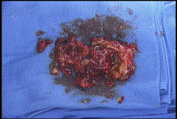
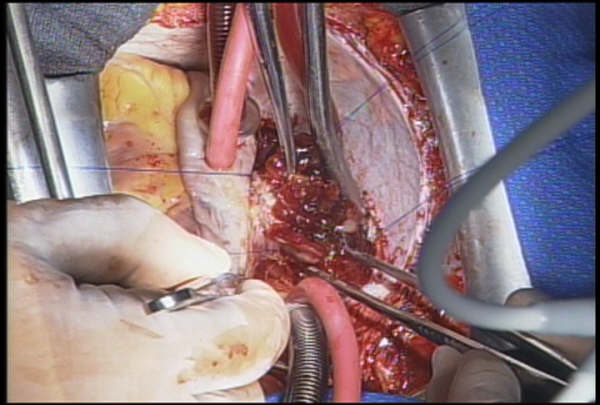
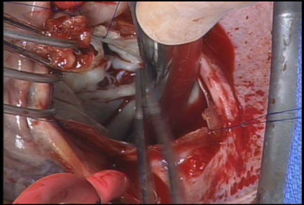
Figures 79a, b and c above are pictures of the specimen of
the left atrial myxoma (video 1) surgically removed from the
left atrium, the left atrium with the tumor partially (Figure
79b) and finally completed removed (Figure 79c).
The
remaining primary cardiac tumor are malignant (meaning that
it is capable of growing wildly in its place of origin The remaining
primary cardiac tumor are malignant (meaning that it is capable
of growing wildly in its place of origin and can metastasize
or spread through the blood and lymph vessels to other parts
of the body), the overwhelming majority of which are sarcomas
(see figure
59). Cardiac sarcomas arise from the right side of the heart
in 25 per cent of reported cases, whereas myxomas arise from
right-sided cardiac structures in only 5 per cent of cases.
Approximately 86 per cent of myxomas develop in the left atrium
(see figures 78a,
78b,
79). An intracavitary tumor that obstructs the right ventricular
outflow tract is about 300 times more likely to be malignant
than benign (figure
80).
Video
1:
Figure 79a: Various views and positions of the myxoma(also
see figure 79b and 79c below)

Figure 79b

Figure 79c

Symptoms
include weight loss, fatigue, fever, anemia, etc. Systemic tumor
embolization occurs in 40-50 per cent, of patients with left
atrial myxoma, with tumor fragments embolizing to the brain,
kidneys and extremities. Symptoms in the above case include
cough, weakness and dyspnea. There was also evidence of pulmonary
hypertension.
Sawaya,
J.I., MD, Dakik, H.A., MD, Angiographic Visualization of an
Atrial Myxoma, The New England Journal of Medicine, Vol 342,
Jan 27-00, p 294-299.
Dennig, K., MD, Lehmann, G., MD, Richter, T., MD, The Angiosarcoma
in the Left Atrium, The New England Journal of Medicine, Vol.
342, Jan 13-00, p 443-444.
Hall, R.J., MD, Cooley, D.A., MD, MCAllister, Jr., H.A., MD,
Frazier, O.H., MD, Neoplastic Heart Disease, Hurst's The Heart,
8th edition, p 2007-2023.
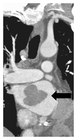
Below is a left atrial myxoma presented by Doctors Ritu Gill, M.D. and Sary F. Aranki, M.D. in the NEW ENGLAND J. MED. 358:7 PAGE728, February 14, 2008. Contrast-enhanced computed tomography confirmed the presence of a left atrial mass (indicated by an arrow in the coronal view of the heart). Pathogical evaluation revealed a benign myxoma.
